

Radiology is broadly thought to be the medical discipline most profoundly reworked by synthetic intelligence. In reality, 76% of all FDA-approved AI algorithms to this point concentrate on synthetic intelligence in medical imaging, a transparent signal of how central this know-how has turn into. These advances are significantly distinguished in pc imaginative and prescient, a subfield of AI centered on decoding visible inputs, which has enabled radiology to evolve right into a extra exact, environment friendly, and scalable self-discipline. Pc imaginative and prescient now powers many core medical imaging instruments, reminiscent of CT and MRI scans, X-rays, and ultrasounds.
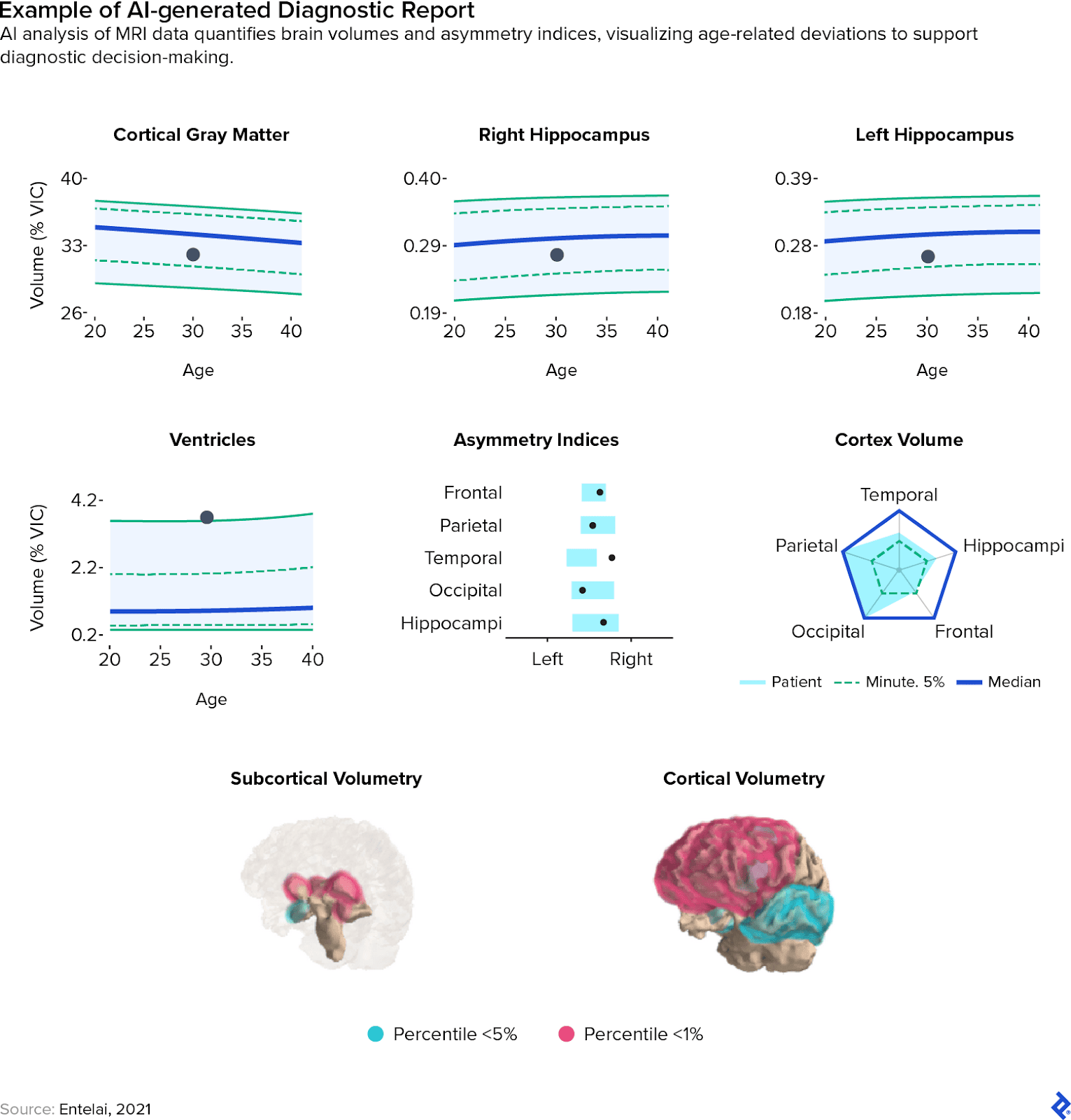
I’ve witnessed this transformation firsthand. As a lead information scientist at Entelai, an AI-and-healthcare startup, I helped develop and deploy a mind MRI evaluation system that integrates straight into the imaging workflows at hospitals. This technique routinely identifies and descriptions demyelinating lesions—areas of nerve fiber injury generally seen in situations like a number of sclerosis—measures mind area volumes, and classifies patterns of atrophy, all whereas feeding outcomes into sufferers’ digital well being data.
The journey of creating this method was not with out its hurdles, however right now, the system is in energetic use throughout a number of imaging facilities in Latin America, with a steadily rising consumer base. This real-world expertise, together with the technical and operational challenges we encountered, frames a lot of what this text explores: how AI in diagnostic imaging is reshaping the sector, from acquisition to analysis.
Overview of AI in Medical Imaging
The fast progress of medical AI imaging is due primarily to 3 key components: the event of neural networks, the supply of enormous quantities of medical imaging information, and the facility of graphics processing models (GPUs) for parallel computing.
At its core, neural networks are machine studying fashions made up of nodes (or “neurons”) which can be interconnected in a layered association. These networks study to course of data by a type of trial and error, adjusting their inside parameters primarily based on suggestions. Convolutional neural networks (CNNs), a subtype of neural networks, are particularly well-suited for pc imaginative and prescient duties resulting from their capacity to extract spatial and hierarchical options from photographs. Every layer identifies a distinct facet of the enter—reminiscent of edges, shapes, or colour gradients—increase a progressively detailed illustration of the picture.
Though CNNs have been the main target of educational analysis for years, their breakthrough efficiency solely turned evident with the emergence of enormous, labeled picture datasets. One landmark instance is ImageNet, a dataset containing tens of millions of annotated photographs organized in accordance with a hierarchical construction. Datasets of this scale reworked pc imaginative and prescient, however coaching giant CNNs on them launched new computational challenges (together with huge energy calls for). That’s the place GPUs turned important. Designed for high-throughput parallel processing, GPUs dramatically lowered coaching instances by dealing with the large variety of operations required to optimize neural networks. This made it doable to coach and take a look at fashions at a a lot sooner tempo.
Developments like these have enabled technological feats that have been unimaginable solely a decade in the past. Face tagging on social media, cell verify scanning, and image-based search have turn into routine elements of each day life. The identical rules energy AI and medical imaging purposes, together with X-rays, CT scans, and ultrasounds. AI imaging methods are being developed to reinforce MRI processes, together with sooner picture reconstruction and superior neuroimaging evaluation, to enhance effectivity and diagnostic capabilities. At Entelai, for instance, we educated CNNs on curated, annotated mind MRI datasets to detect lesions and estimate mind area volumes. GPU acceleration was essential, significantly through the iterative strategy of enhancing segmentation accuracy.
AI medical imaging provides vital advantages: greater diagnostic accuracy, sooner picture processing, and decrease general prices. Nevertheless, the sensitivity of healthcare environments implies that automation have to be approached with warning. Challenges and moral issues—reminiscent of transparency, bias mitigation, and medical validation—have to be built-in into each stage of product improvement.
High 3 Advantages of Synthetic Intelligence in Medical Imaging
Synthetic intelligence in medical imaging helps radiologists work sooner and extra precisely, whereas lowering fatigue. This strengthens a workforce beneath vital pressure, as the sector presently faces a international expertise scarcity. A number of components contribute to this hole, together with elevated burnout amongst professionals and a surge in demand pushed by an growing older international inhabitants. Members of the scientific and medical communities are more and more seeking to imaging AI as a manner to assist alleviate this bottleneck by enabling radiologists to work extra effectively and with fewer fatigue-related errors. Under are three key methods AI medical imaging is shifting the sector towards a extra scalable and resilient future.
1. AI Imaging Is a Quantitative Complement to Qualitative Medical Imaging Evaluation
Radiologists depend on years of coaching and expertise to interpret medical photographs and draw significant conclusions. The human eye excels at detecting nuanced patterns and integrating contextual data—reminiscent of affected person medical historical past—into diagnostic reasoning. Nevertheless, AI-powered pc imaginative and prescient techniques are uniquely suited to extracting exact measurements and conducting large-scale quantitative analyses that will be time-prohibitive for human specialists. As an example, delineating and calculating the amount of particular mind areas from an MRI scan is often too labor-intensive to carry out manually, however picture evaluation AI instruments can full this activity quickly and precisely.
In apply, at Entelai, we noticed that AI-generated quantitative outputs helped radiologists validate their medical imaging evaluation extra confidently and constantly, even in fast-paced medical environments. Reasonably than changing professional judgment, AI imaging augments it with goal information and reproducible measurements.
2. AI Medical Picture Evaluation Reduces Error Charges and Improves Response Instances
Fatigue from prolonged shifts will increase the danger of diagnostic errors. AI medical imaging techniques can act as dependable “second opinions” for radiologists, flagging potential oversights and prompting medical specialists to overview ambiguous or high-risk findings. As well as, AI-powered triage instruments can assist optimize workflows by prioritizing pressing or complicated circumstances for overview early in a shift, when clinicians are most alert.
These techniques can analyze variables reminiscent of shift schedules and historic efficiency information to assign circumstances extra strategically. Whereas approaches differ by implementation, triage-driven workflows have proven promising ends in lowering diagnostic delays. For instance, in a examine centered on chest X-rays, AI prioritization lowered the typical turnaround time for picture experiences from 11.2 days to simply 2.7 days. Related positive factors could also be achievable throughout different imaging modalities as AI instruments mature, translating to faster diagnoses and earlier interventions for sufferers.
3. Diminished Prices and Spherical-the-Clock Availability
By automating repetitive duties and streamlining picture evaluation, AI in diagnostic imaging has the potential to scale back operational prices. These efficiencies could make diagnostic companies extra inexpensive for healthcare techniques and extra accessible to sufferers. Not like human radiologists, AI techniques don’t require relaxation and might present uninterrupted help to care groups no matter time or staffing constraints. This 24/7 functionality is especially beneficial in distant or underserved areas, the place entry to professional interpretation is commonly restricted.
3 Key Challenges of Utilizing AI in Diagnostic Imaging
Regardless of its vital advantages, deploying AI for diagnostic functions has moral and technological challenges that have to be fastidiously addressed. These should not summary points however moderately ones with vital regulatory implications, particularly as worldwide frameworks reminiscent of GDPR and the EU AI Act more and more set necessities for issues like transparency and accountability. Under are three key hurdles going through AI in medical imaging right now.
1. Dataset Bias
In curating a coaching dataset throughout clinics in Latin America, I noticed firsthand how complicated and important this step will be. A machine studying mannequin is solely as dependable as the info it’s educated on. When datasets lack range or fail to mirror the populations they’re meant to serve, the ensuing fashions can carry ahead these biases. Analysis has demonstrated that this could result in inaccurate diagnoses for underrepresented teams, exacerbating well being disparities.
To keep away from this, datasets must be constructed with deliberate consideration to demographic and medical range. Methods like dataset rebalancing, information augmentation, and mannequin recalibration can assist mitigate bias, however these efforts have to be a part of a broader, ongoing dedication to moral AI improvement. Our work at Entelai required a collaborative partnership with quite a few clinics, partaking them within the creation of a complete dataset, with the consent of particular person sufferers who agreed to contribute to the info pool.
2. Mannequin Generalization
For synthetic intelligence in medical imaging to be clinically helpful throughout totally different healthcare settings, it should reliably carry out on new information from assorted imaging tools and affected person populations. This capacity, often called generalization, is without doubt one of the key challenges in creating diagnostic AI techniques. In sensible phrases, generalization refers to a mannequin’s capability to use what it has discovered from one dataset to a separate, beforehand unseen set of knowledge.
For instance, a mannequin educated on mind scans from one producer’s MRI machines could not carry out as properly when utilized to pictures from a distinct vendor, resulting from variations in decision, distinction, or imaging protocols. These discrepancies can cut back diagnostic accuracy and undermine medical belief. Consequently, constructing strong, generalizable fashions requires not solely various coaching information but additionally energetic validation throughout a spread of sources.
We encountered this problem throughout our deployment pipeline at Entelai. Variations in imaging tools and acquisition settings throughout clinics sometimes led to drops in system efficiency. To handle this, we applied a devoted monitoring framework, vigilantly overseen by our high quality assurance workforce, to trace the combination of every new clinic. When vital efficiency degradation was detected, our workforce responded by fine-tuning current fashions or creating devoted ones calibrated to the precise information distribution of the brand new atmosphere.
This expertise underscored the significance of reside monitoring, a vital part of accountable AI deployment. Monitoring helps detect underperformance early, permitting groups to intervene earlier than inaccuracies attain sufferers or clinicians. In dynamic medical settings, the place real-world situations typically differ from coaching eventualities, adaptability and oversight are essential to sustaining mannequin efficiency.
3. Lack of Explainability
AI medical imaging fashions typically operate as black packing containers, that means their decision-making processes will be opaque, even to the builders who constructed them. Whereas these techniques could ship correct outcomes, their lack of interpretability presents a barrier to medical belief and adoption. Clinicians and sufferers alike want to grasp the rationale behind a analysis and never simply settle for it at face worth.
To deal with this, researchers have developed a wide range of explainability methods, usually falling into three classes:
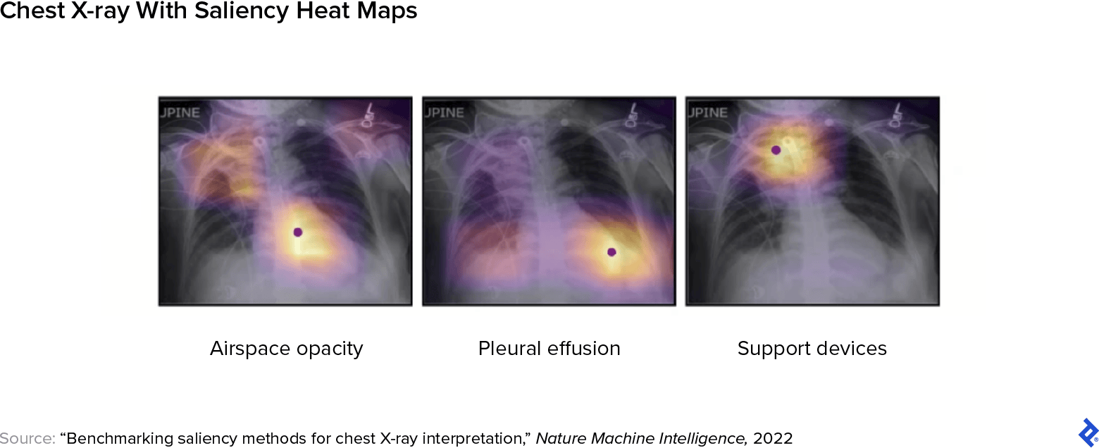
- Visible methods, reminiscent of saliency maps, spotlight the picture areas that almost all influenced the mannequin’s output, permitting clinicians to see the place the mannequin “seemed” when forming its conclusion. For instance, a saliency map superimposed on a CT scan can assist pinpoint the world the place the mannequin detected an anomaly.
- Textual methods generate concise explanations that describe the mannequin’s reasoning in plain language. These are sometimes applied as choice help textual content, providing clinicians a written abstract of the logic behind the output.
- Statistical methods present structured information—reminiscent of characteristic significance scores or mannequin comparisons—that make clear how predictions have been made. Function significance evaluation identifies which picture elements most impacted the result, whereas mannequin comparability permits groups to judge how totally different fashions carry out on the identical dataset.
Explainability is a quickly evolving discipline, and enhancing transparency will likely be important for constructing belief and making certain protected, accountable deployment of AI in diagnostic workflows. The EU’s AI Act from 2024 contains an express regulatory requirement pertaining to transparency and interpretability in high-risk purposes. By August 2026, these techniques have to be designed in order that healthcare suppliers can interpret AI outputs and use them appropriately, supported by clear data on efficiency limitations and identified dangers.
How Correct Is AI in Medical Imaging?
Analysis exhibits that in some purposes, AI techniques carry out on par with—and even exceed—the accuracy of professional radiologists. In mammography screening, for instance, a big population-based examine discovered that AI matched the efficiency of double studying—a quality-control apply through which two impartial radiologists overview every scan—whereas additionally lowering workload by 44%. In a separate examine on breast ultrasounds, AI techniques lowered false positives by 37.3% and pointless biopsies by 27.8%.
Nonetheless, accuracy relies upon closely on the use case and dataset high quality. Researchers have discovered that fashions educated in managed analysis environments can underperform when deployed in real-world medical settings, the place affected person populations and picture high quality differ. This underscores the significance of human-AI collaboration in medical imaging: AI must be thought to be a complement to human experience, pairing computational velocity and sample recognition with on-the-ground medical judgment. Ongoing efficiency validation and human oversight are important to make sure dependable, context-appropriate outcomes.
4 Steps to Modernize Radiological Workflows With AI Imaging
Each stage of the medical imaging workflow presents alternatives for enchancment by AI. AI fashions are utilized to detect early indicators of ailments reminiscent of breast most cancers, lung most cancers, tuberculosis, and cardiovascular dangers. From the second a picture is acquired to the ultimate reporting part, AI applied sciences are serving to radiologists work with higher effectivity and accuracy. Under is a step-by-step overview of how AI is being built-in into the radiological pipeline.
Step 1: Picture Acquisition
Step one in any imaging workflow is buying a clear, high-quality picture. Errors launched right here can cascade all through the pipeline, resulting in elevated radiation publicity for sufferers or diagnostic delays. AI instruments can now help with minimizing these dangers and enhancing acquisition effectivity in the most typical medical imaging use circumstances.
- CT: Programs like Radiology Sensible Assistant help automated affected person positioning, aligning the physique accurately inside the scanner to make sure the area of curiosity is centered and inside the discipline of view. These instruments additionally carry out error flagging, figuring out points reminiscent of movement artifacts in actual time. This reduces pointless radiation publicity and improves scan effectivity.
- MRI: Siemens Healthineers applies deep studying to speed up picture acquisition and enhance picture reconstruction, a course of that transforms uncooked sign information into clear, high-resolution photographs. AI also can automate slice positioning, deciding the place within the physique to amass photographs and suggesting the suitable scan protocol primarily based on medical context.
- Fluoroscopy: FluoroShield allows automated collimation, which dynamically adjusts the scale and form of the X-ray beam throughout procedures. This minimizes pointless radiation publicity to surrounding tissue and improves picture concentrate on the world of curiosity, which is essential in dynamic research of the gastrointestinal or cardiovascular techniques.
- Ultrasound: AI-assisted ultrasound instruments acknowledge anatomical landmarks in actual time, guiding the sonographer to the proper imaging planes and automating organ identification. That is particularly useful in point-of-care settings, the place scan high quality can considerably have an effect on analysis.
Step 2: Picture Preprocessing
As soon as a picture is captured, it have to be reworked from uncooked information right into a clinically usable format. This stage, often called preprocessing, entails noise discount, artifact correction, and picture reconstruction to reinforce consistency and diagnostic high quality. AI applied sciences have been particularly impactful right here, enabling sooner and extra correct processing, even when scan situations are lower than splendid.
- CT: Instruments like TrueFidelity use deep studying to reconstruct photographs from low-dose scans, preserving high-quality anatomical element whereas minimizing radiation publicity. By studying from high-quality examples, these fashions can cut back artifacts and enhance distinction, serving to radiologists interpret refined findings with higher confidence.
- MRI: AI is utilized to speed up MRI scan instances and enhance reconstruction constancy, even with undersampled information. Deep studying fashions can get well lacking spatial data and cut back noise, producing clearer photographs with sharper decision, which is particularly beneficial for detecting small lesions or refined structural modifications.
Regardless of the constructive implications for these use circumstances, it’s essential to notice that each one picture enhancement methods carry some threat of introducing artifacts or over-smoothing areas, doubtlessly masking abnormalities or producing particulars that weren’t current within the unique scan. Sturdy validation and medical oversight stay important to make sure diagnostic reliability.
Step 3: Picture Evaluation and Interpretation
As soon as a picture is preprocessed and prepared for interpretation, the subsequent step is evaluation, which is arguably the part the place the mixture of AI and medical imaging has essentially the most thrilling implications. Right here, AI fashions help with figuring out abnormalities and classifying medical situations, and might automate routine duties like picture segmentation, permitting radiologists to concentrate on extra complicated diagnostic challenges. These duties type the premise for a lot of medical choices and are sometimes essentially the most labor-intensive for radiologists.
- Detection: This entails routinely figuring out the presence and placement of options reminiscent of tumors, fractures, or nodules inside a picture. AI medical picture evaluation fashions are educated to acknowledge particular patterns, just like the density or form of an irregular mass, and might flag areas of curiosity with bounding packing containers or markers. Detection instruments can speed up analysis time and cut back the danger of overlooking refined or incidental findings.
- Segmentation: On this step, AI techniques isolate anatomical buildings or abnormalities by dividing a picture into labeled areas. For instance, segmentation fashions can define mind ventricles, delineate tumor margins, or separate lung lobes, typically all the way down to the pixel degree. This allows exact measurements, volumetric evaluation, and 3D reconstructions, that are essential for therapy planning, surgical navigation, and illness monitoring.
- Classification: After detection and segmentation, AI can assign diagnostic labels to findings, reminiscent of figuring out whether or not a lesion is benign or malignant, or classifying tissue modifications in step with illness development. Classification fashions typically incorporate probabilistic scoring, giving clinicians a confidence degree that may information additional investigation or motion.
The detection, segmentation, and classification capabilities underpin a variety of medical purposes for synthetic intelligence in medical imaging:
- X-ray: AI instruments for chest X-rays can detect situations like pneumonia, pleural effusion, or cardiomegaly, typically with accuracy corresponding to human radiologists. In mammography, AI is being deployed as a second reader to establish refined indicators of breast most cancers and cut back false negatives. These techniques assist triage high-risk circumstances and supply choice help in settings with restricted radiology experience.
- CT: Through the COVID-19 pandemic, AI-driven CT evaluation emerged as a strong software for detecting pulmonary hallmarks of the illness, enabling clinicians to triage sufferers in high-pace, high-volume medical settings. These instruments additionally supported monitoring of illness severity and evaluation of long-term lung modifications, which frequently persist months after restoration. Individually, in dental imaging, absolutely automated AI techniques have been built-in with cone-beam CT (CBCT) to delineate dental buildings, attaining segmentation accuracy corresponding to that of skilled radiologists, whereas working as much as 500 instances sooner.
- MRI: In neuroimaging, AI instruments like FastSurfer use deep neural networks to speed up mind segmentation workflows by automating the identification of areas affected by situations reminiscent of a number of sclerosis. In the meantime, the AI system we developed at Entelai enabled automated segmentation of cortical and subcortical mind areas, supporting medical analysis and the longitudinal monitoring of neurodegeneration.
- Ultrasound: AI applied sciences are getting used to enhance consistency and diagnostic accuracy in a wide range of ultrasound purposes, together with breast imaging and echocardiography. Specifically, deep studying can information picture acquisition and automate interpretation, lowering reliance on operator experience and increasing entry to high-quality care, even in resource-limited or distant settings.
Step 4: Reporting and Scientific Communication
The ultimate step within the radiological workflow is the era of structured experiences, which synthesize findings into actionable insights for referring physicians. These experiences are extremely specialised, typically dense with medical terminology, and should steadiness precision with effectivity, particularly in high-volume settings. Historically, radiologists dictate or manually enter findings, however this course of will be time-consuming and liable to inconsistencies.
AI is starting to remodel this part by supporting each report era and comprehension. Pure language processing (NLP) instruments can routinely extract key data from picture analyses and draft preliminary experiences, lowering documentation burden and standardizing terminology. Whereas not infallible, these techniques can help consistency and velocity when used alongside professional overview. Superior techniques are additionally exploring report simplification, translating technical findings into accessible language for nonspecialist physicians or sufferers. As these capabilities evolve, they maintain the potential to streamline communication and enhance continuity of care throughout medical groups.
Understanding the Constructing Blocks of AI in Diagnostic Imaging
AI fashions differ primarily based on the kind of enter they course of—reminiscent of photographs, textual content, audio, or sensor information—and the type of output they’re designed to supply. Many medical instruments mix a number of fashions right into a pipeline, and even into multimodal techniques that combine imaginative and prescient, language, and real-time sensor suggestions.
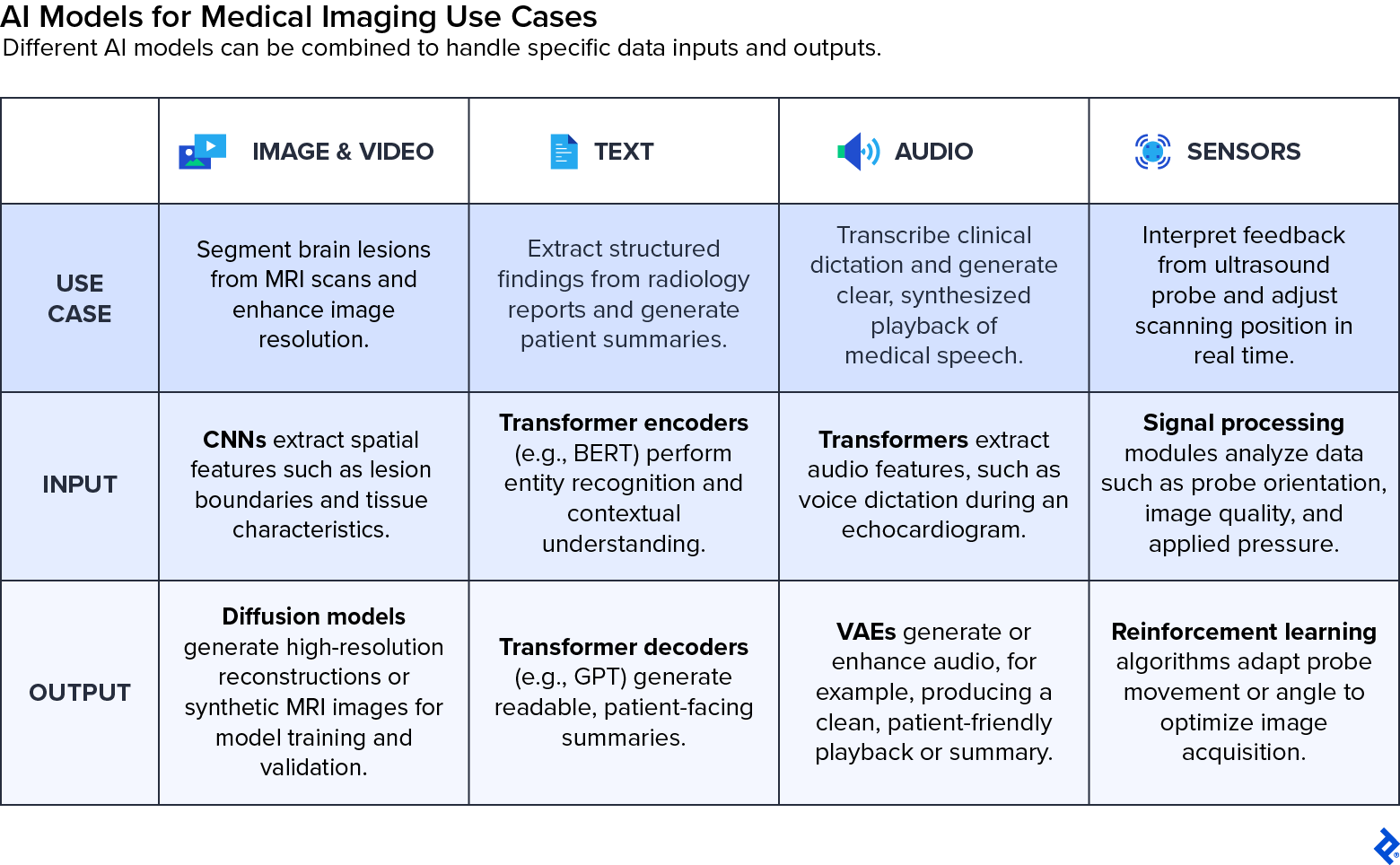
Under is an outline of the primary forms of medical imaging AI fashions and the way they’re used:
- Picture and video fashions are on the core of radiology AI, together with the system we developed at Entelai. CNNs are generally used for classification and segmentation duties, reminiscent of detecting lung nodules or measuring organ volumes. Extra superior architectures like imaginative and prescient transformers seize broader spatial context, whereas generative fashions, reminiscent of generative adversarial networks (GANs), diffusion networks, and variational autoencoders (VAEs), are more and more used for picture enhancement, artificial information era, and magnificence switch.
- Textual content fashions assist construction and interpret the written elements of radiology workflows. Transformer-based fashions like GPT and BERT (brief for bidirectional encoder representations from transformers) can summarize experiences, extract structured findings, and even translate technical imaging descriptions into patient-friendly language. Different fashions use phrase embeddings to seize the that means of medical phrases primarily based on context, or lengthy short-term reminiscence networks (LSTMs) to course of textual content sequences reminiscent of radiology notes over time. It’s essential to notice that textual content fashions can sometimes generate hallucinations, so healthcare suppliers should at all times overview their outputs fastidiously to make sure medical accuracy.
- Audio fashions are utilized in medical environments for sign processing, speech recognition, and voice command performance. These embody recurrent neural networks (RNNs), LSTMs, and transformers that energy real-time transcription instruments, enabling radiologists to dictate findings hands-free. Along with transcription, conventional speech synthesis methods and variational autoencoders are used to create or improve medical audio, reminiscent of producing natural-sounding speech or filtering out noise from recordings.
- Sensor and actuator fashions interpret real-time enter from bodily gadgets, reminiscent of ultrasound probes or robotic devices, and generate output alerts that information movement or positioning. These fashions depend on sign processing, management concept, and reinforcement studying to repeatedly regulate actions primarily based on sensor suggestions, enhancing precision and lowering the necessity for guide correction throughout procedures.
As medical imaging AI techniques develop extra subtle, these mannequin varieties are more and more built-in throughout modalities. For instance, a multimodal platform would possibly mix mind MRI segmentation, medical textual content evaluation, and longitudinal monitoring to help extra holistic assessments of neurodegenerative ailments. This convergence is opening the door to diagnostic techniques that synthesize various medical information streams to ship broader patient-centered perception.
What Are the Future Tendencies of AI in Medical Imaging?
The present wave of medical imaging AI has been pushed largely by advances in pc imaginative and prescient, with further positive factors from pure language processing for report era and structured documentation. However a brand new chapter is starting—one centered on multimodal AI techniques that may combine a wide range of inputs, together with medical photographs, medical textual content, sensor information, and affected person historical past, right into a unified diagnostic mannequin. The developments level towards the arrival of generalist medical AI fashions able to remodeling the whole diagnostic course of.
These next-generation techniques intention to maneuver past analyzing a single scan in isolation. As an alternative, they will synthesize data throughout modalities and time, factoring in prior imaging, lab outcomes, and doctor notes to generate extra complete and clinically helpful experiences. This shift mirrors how human clinicians purpose, drawing from a number of information factors to tell judgment moderately than relying solely on one picture or take a look at.
Throughout my time at Entelai, we took foundational steps on this route. We built-in automated mind MRI evaluation with digital well being data to assist radiologists interpret new findings within the context of quantitative tendencies over time. As these capabilities evolve, they provide a glimpse of how AI may transfer past picture interpretation to start supporting broader medical reasoning.
To appreciate this potential, technical progress have to be matched by cautious validation and moral deployment. The deployment of AI in medical imaging is influenced by regulatory hurdles and the necessity for clear algorithms that construct belief and guarantee accountability. Points like explainability, regulatory approval, dataset bias, and the necessity for high-quality, labeled information to make sure correct and equitable outcomes will form how shortly and responsibly these techniques attain widespread adoption. Academic initiatives and summits are exploring these moral issues in addition to the authorized features and coaching necessities for integrating AI into radiology practices.
Integrations should emphasize the significance of human experience, the place synthetic intelligence in medical imaging aids however doesn’t change radiologists, enhancing diagnostic accuracy and effectivity whereas preserving high-quality affected person care and human medical judgment. If performed proper, this subsequent part of AI imaging may rework diagnostics from a static snapshot right into a repeatedly studying, decision-support ecosystem that delivers richer and extra personalised care at scale.
The technical content material offered on this article was reviewed by Roman Vlasov.